Brain Classification
The brain has a complex anatomy. In particular, its cortical surface is a complex manifold containing many convex structures, called gyri, and concave structures, called sulci. These structures construct complex patterns which convey important information about the brain anatomy and, possibly, function. Several applications require the robust detection of sulci and gyri, for example for comparison of different brain anatomies, or registration of the same brain anatomy measured at different moments.
Traditionally, sulci and gyri (and similar structures in other anatomical datasets) have been detected using measurements of the surface curvature minima and maxima respectively. Although many effective methods exist in this area, we are interested to study if such structures can be detected without curvature estimation, for two reasons:
- curvature estimations involve differentiation and, as such, are inherently delicate on noisy or poorly sampled datasets
- obtaining the same results with a curvature-free method opens the way for curvature-free methods in many other areas of research
Skeleton-based classifiers
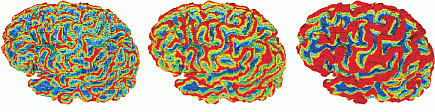
We have developed a so-called surface classifier that discriminates between convex and concave surface regions using a fully curvature-free method. Instead, we use 3D surface skeletons. Using a simplified skeleton, we detect all shape (brain surface) points corresponding to its feature points. This creates gaps in the surface corresponding precisely to the gyri (convex features) centers. Using the shape background skeleton, we detect the sulci centers. The geodesic distance on the surface to these centers is our classifier. Progressively simplified skeletons give us classifiers at different scales of the curvature.
The image below shows a classifier (red=gyri, blue=sulci) for three different scales computed with our method. Note the sharp separation of gyri from sulci.
Comparison
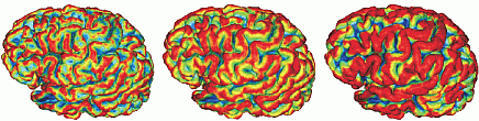
The image below shows the same brain as in the prior image, this time classified using the well-known curvature-based classifier of Taubin. The classification is noisier, and the separation between gyri and sulci is harder to see.
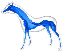
Brain structures
Sulci and gyri do not follow random patterns, but are inherently related to brain functions. As a first step to understand the anatomical patterns they represent, we investigate the use of skeletons. For this, we compute the 3D surface and brain skeletons and next segment these, as described here. The image below shows such a segmentation. Given the stability of our multiscale skeletons to small surface changes, we believe it is possible to use such skeletons as a first step in comparing, matching, or registering different brain anatomies.
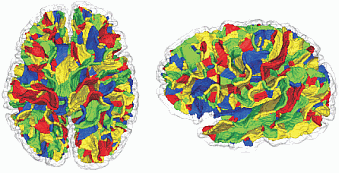
Segmented surface skeleton of a brain

Segmented curve skeleton of the same brain
Publications
Robust Classification and Analysis of Anatomical Surfaces Using 3D Skeletons D. Reniers, A. Jalba, A. Telea. Proc. VCBM'08, Eurographics, 2008
Software
The brain classification is implemented in the software described here.