Visual Analytics for Engineering Skin Lesion Classifiers
MAaglinant melanoma are one of the most aggressive cancer forms; the most aggressive skin cancer type; and has a rapid incidence in the last two decades. Detecting melanoma (or naevi prone to transform into melanoma) in an early stage significantly increases the chances of treatment and next of prognosis. However, this requires time-consuming examination by dermatologists and/or intrusive bioptic procedures.
Visual classification
We have developed and end-to-end machine learning system able to discriminate melanoma from other similar skin lesions, such as moles, based on a visual analytics approach. The system works as follows:
- Over 600 image features (common in generic image classification) are extracted from images collected from a variety of sources (in-the-field use of various dermatoscopic devices such as HandyScope; in-polyclinic collected images by medical specialists; and images from well-known medical databases).
- Several classifiers are next trained and tested on these data, including KNN, LVQ, LogReg, RFC, and DecTrees.
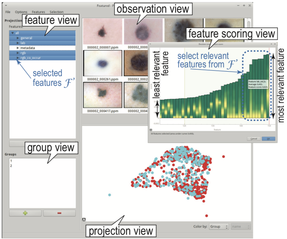
- Classification results and ground-truth are visually compared using dimensionality-reduction projections
- The specialist can then eliminate groups of features which appear to be redundant and/or not instrumental for the ground-truth class separation, to simplify the classifier operation and/or increase its accuracy. The new classifier is re-designed, trained, and visualized on-the-fly
- Iterating the last two steps allow fine-tuning an optimal set of features leading to a fast-to-execute, simple-to-implement, and high-accuracy classifier.
Results
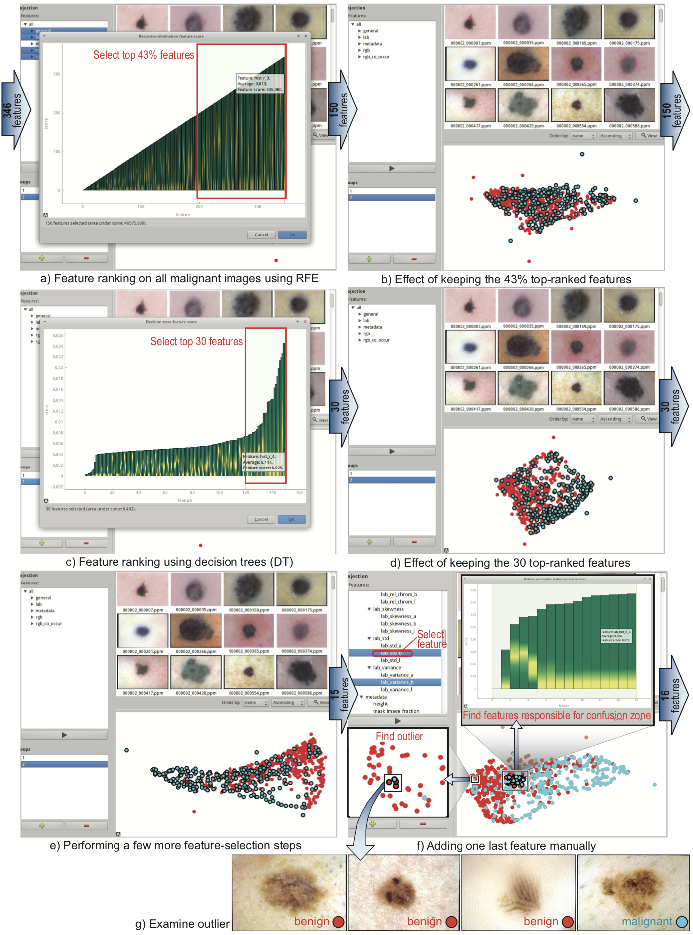
We tested our system on several real-world datasets, including established benchmarks (over 2000 images, with ground-truth labels given by dermatologists). The image below shows six steps of our iterative procedure which allowed us to reduce the number of extracted and used features from 346 to only 14 features, and increase its accuracy by 2%. The entire visual exploration-and-feature-selection process took under 10 minutes.
Implementation
The system is implemented using our featured high-dimensional data exploration tool.
Acknowledgments
The project (MelanoImage) was partially sponsored by a grant from ANCS Romania (2011-2014)
Publications
Interactive Image Feature Selection Aided by Dimensionality Reduction P. Rauber, R. da Silva, S. Feringa, M. Celebi, A. Falcao, A. Telea. Proc. EuroVA, 2015
Comparison of Features used in Automatic Skin Lesion Classification S. Feringa, MSc thesis, Univ. of Groningen, 2015